Medical imaging has transformed the way neurological conditions are diagnosed and managed. From detecting early-stage tumors to identifying the precise location of a stroke, neuroradiology sits at the intersection of advanced technology and clinical precision. Yet many patients remain unfamiliar with what a neuroradiologist actually does, when a referral is appropriate, and how to prepare for the diagnostic process. This article breaks down the essentials of neuroradiology, what to expect during imaging procedures, and how to find the right specialist for your needs.
What Is Neuroradiology and Why Does It Matter?
Neuroradiology is a subspecialty of radiology focused exclusively on diagnosing and, in some cases, treating conditions affecting the brain, spine, head, neck, and peripheral nervous system. Unlike a general radiologist who interprets a broad range of imaging studies, a neuroradiologist undergoes additional fellowship training specifically in neurological imaging modalities. This depth of expertise allows them to detect subtle abnormalities that might be missed in a general reading — a distinction that can be life-changing for patients with complex or rare conditions.
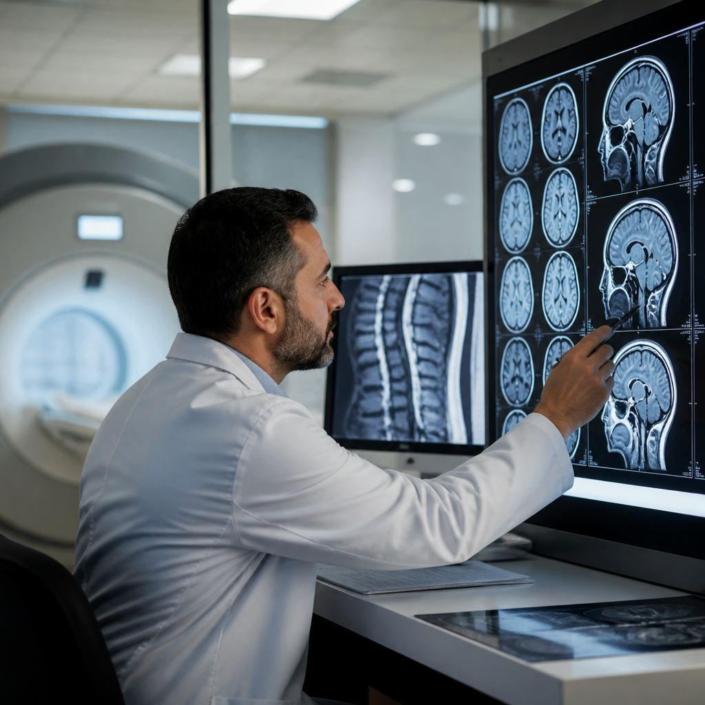
The tools of the trade include magnetic resonance imaging (MRI), computed tomography (CT), positron emission tomography (PET), and interventional procedures such as cerebral angiography. Each modality serves a different diagnostic purpose, and a skilled neuroradiologist knows precisely which technique — or combination of techniques — will yield the most clinically useful information for a given patient presentation.
Common Conditions Diagnosed Through Neuroradiology
Neuroradiological imaging is used to evaluate a wide spectrum of conditions. Some of the most frequently encountered include:
Stroke and Cerebrovascular Disease
Time is brain tissue when it comes to stroke. Neuroradiologists play a critical role in rapidly interpreting CT and MRI scans to determine whether a stroke is ischemic or hemorrhagic, which directly dictates the treatment pathway. Advanced perfusion imaging can even identify salvageable brain tissue, guiding decisions about thrombectomy or thrombolysis.
Brain Tumors and Lesions
Identifying a brain tumor is only the beginning. Neuroradiologists characterize lesions by their size, location, vascularity, and relationship to surrounding structures. Spectroscopy and perfusion MRI can help differentiate between tumor types and distinguish recurrent tumor from treatment-related changes — information that is invaluable for neurosurgeons and oncologists planning care.
Spine Disorders
From herniated discs and spinal stenosis to demyelinating diseases and spinal cord tumors, the spine presents a complex imaging landscape. Neuroradiologists interpret these studies with an understanding of both the structural anatomy and the clinical syndromes they produce, ensuring that radiological findings are translated into actionable clinical guidance.
Multiple Sclerosis and Demyelinating Diseases
MS diagnosis relies heavily on MRI criteria, and neuroradiologists are trained to identify characteristic plaques, assess lesion burden, and monitor disease progression over time. Standardized imaging protocols ensure consistency in follow-up studies, which is essential for tracking treatment response.
The Role of Interventional Neuroradiology
Beyond diagnosis, a subset of neuroradiologists specializes in minimally invasive procedures performed under imaging guidance. These interventional techniques include cerebral angiography, coiling of brain aneurysms, mechanical thrombectomy for acute stroke, and vertebroplasty for spinal compression fractures. These procedures often offer patients a less invasive alternative to open surgery, with shorter recovery times and reduced risk. The precision required for these interventions underscores why subspecialty training in neuroradiology is so critical.
How to Prepare for a Neuroradiological Procedure
Preparation varies depending on the type of imaging study ordered, but there are universal steps every patient should take before any diagnostic procedure. Reviewing these practical steps to prepare for medical procedures can help patients feel more confident and reduce anxiety on the day of their appointment. Key considerations include disclosing all medications and supplements to your care team, fasting requirements for contrast-enhanced studies, and informing staff of any implanted devices such as pacemakers or cochlear implants that may be contraindicated with MRI.
Patients undergoing contrast-enhanced MRI or CT should also be aware of potential allergic reactions and kidney function considerations, particularly if they have pre-existing renal disease. Your referring physician and the radiology team will guide you through any specific preparation requirements, but arriving informed and prepared makes the entire process smoother for everyone involved.
Building a Sustainable Diagnostic Pathway
For clinicians managing patients with complex neurological conditions, establishing a reliable and efficient diagnostic pathway is essential — not just for patient outcomes, but for the sustainability of the practice itself. Understanding how to grow a medical practice without burning out is increasingly relevant in specialties like neuroradiology, where the cognitive demands are high and the margin for error is narrow. Integrating streamlined referral systems, clear communication protocols between neurologists and radiologists, and patient education resources can significantly reduce friction in the diagnostic process while improving overall care quality.
Finding Expert Neuroradiological Care in the UAE
Access to subspecialty neuroradiology expertise has historically been limited in many parts of the Middle East, but that landscape has changed significantly in recent years. Patients in the region now have access to internationally trained specialists operating within world-class facilities. Consulting a qualified neuroradiologist in abu dhabi at Cleveland Clinic Abu Dhabi means benefiting from a team that applies the same diagnostic standards and imaging protocols used at one of the most respected medical institutions in the world. This level of expertise is particularly important for patients with complex or rare neurological conditions who require precise, nuanced interpretation of their imaging studies.
Cleveland Clinic Abu Dhabi: A Center of Neuroradiological Excellence
Cleveland Clinic Abu Dhabi’s neuroradiology program is staffed by fellowship-trained specialists who bring deep expertise across the full spectrum of neurological imaging. The department is equipped with state-of-the-art MRI, CT, and interventional suites, enabling comprehensive evaluation and treatment of conditions ranging from common spine disorders to rare vascular malformations. The multidisciplinary model — where neuroradiologists collaborate directly with neurologists, neurosurgeons, and oncologists — ensures that imaging findings are interpreted within the full clinical context of each patient’s condition. This integrated approach reduces diagnostic delays and supports more precise, personalized treatment planning.
Conclusion: Precision Imaging as the Foundation of Neurological Care
Neuroradiology is far more than a technical service — it is a clinical discipline that directly shapes the trajectory of neurological care. Whether you are a patient experiencing unexplained neurological symptoms, a caregiver navigating a loved one’s diagnosis, or a clinician seeking a reliable imaging partner, understanding the scope and value of neuroradiology is essential. With the right specialist, the right technology, and the right preparation, diagnostic imaging becomes a powerful tool for clarity, precision, and ultimately, better health outcomes.